
Dr Suvir Gupta -
About Me
Dr Suvir Gupta -
MBBS
Cardiologist
10 Years Experience
Biography
My Clinic
OPD Hours
| Monday | Morning | 10:00 AM | - | 02:00 PM |
| Evening | 06:00 PM | - | 09:00 PM | |
| Tuesday | Morning | 10:00 AM | - | 02:00 PM |
| Evening | 06:00 PM | - | 09:00 PM | |
| Wednesday | Morning | 10:00 AM | - | 02:00 PM |
| Evening | 06:00 PM | - | 09:00 PM | |
| Thursday | Morning | 10:00 AM | - | 02:00 PM |
| Evening | 06:00 PM | - | 09:00 PM | |
| Friday | Morning | 10:00 AM | - | 02:00 PM |
| Evening | 06:00 PM | - | 09:00 PM | |
| Saturday | Morning | 10:00 AM | - | 02:00 PM |
| Evening | 06:00 PM | - | 09:00 PM | |
| Sunday | Morning | 10:00 AM | - | 02:00 PM |
| Evening | 06:00 PM | - | 09:00 PM |
Treatments
Specialised care and treatments offered
CARDIAC REHABILITATION

A structured program combining exercise training, education, and counseling improves recovery following cardiac events like myocardial infarction or procedures such as angioplasty.
IMPLANTABLE LOOP RECORDER
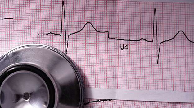
Certain patients suffer from sporadic fainting spells or palpitations that elude diagnosis via conventional tests. An implantable loop recorder offers long-term monitoring of cardiac rhythms directly inside the body.
STRESS TESTING (EXERCISE STRESS TEST)
The heart’s response to physical exertion reveals important clues about underlying coronary artery disease. Stress testing evaluates cardiac performance during controlled exercise conditions.
- SIGNALS FOR USE: Recommended for people with chest discomfort or those at risk of cardiovascular problems seeking diagnosis or clearance before activity.
- TASK DETAILS: Walking on a treadmill or pedaling stationary bike while monitoring ECG, blood pressure, and symptoms closely detects abnormalities induced by stress.
- DYNAMICS OF RESULTS: Decreased exercise tolerance or abnormal ECG changes indicate possible ischemia requiring further investigation or intervention.
- SPECIAL CONSIDERATIONS: Some individuals unable to exercise undergo pharmacologic stress testing using medications that simulate exercise effects on the heart.
ECHOCARDIOGRAPHY
A clear evaluation of heart structure and function is achieved through echocardiography, an ultrasound-based imaging technique used widely in cardiology clinics.
- PURPOSE: It assesses chamber sizes, valve function, wall motion abnormalities, and fluid accumulation around the heart.
- SIGNIFICANCE: Conditions like valve diseases, cardiomyopathies, and congenital defects are identified early with this safe and painless test.
- METHOD: A transducer emits sound waves that reflect off cardiac tissues producing images displayed on a monitor in real-time.
- EFFECTIVENESS: This examination guides treatment decisions—ranging from medication management to surgical repair when needed.
The test provides comprehensive information about heart health without exposure to radiation and has no downtime for patients afterward.
HOLTER MONITORING
Detecting irregular heart rhythms over time can be challenging during brief clinic visits. Holter monitoring offers a continuous recording of the heart’s electrical activity for 24-48 hours or longer.
- Indications: Patients experiencing palpitations, unexplained dizziness, or fainting may benefit from this test.
- Procedure details: A portable device with electrodes attached to the chest records ECG signals throughout daily activities and sleep.
- Analysis: Data are reviewed to identify arrhythmias like atrial fibrillation, premature beats, or pauses in heartbeat.
- Treatment guidance: Results help tailor medication adjustments or decide on further interventions such as pacemaker implantation.
This non-invasive tool provides invaluable insight into intermittent cardiac issues that might otherwise go undetected during routine exams.
CORONARY ANGIOPLASTY
When arteries supplying the heart become narrowed or blocked, restoring blood flow is essential to prevent heart attacks. Coronary angioplasty is a minimally invasive procedure designed to open these obstructed vessels.
- Causes and symptoms: Blockages often result from atherosclerosis, leading to chest pain (angina), shortness of breath, or fatigue.
- Diagnostic approach: Techniques like coronary angiography visualize blockages using contrast dye and X-rays.
- Treatment method: A catheter with a balloon is threaded into the artery; inflating it compresses plaque against the wall. Often, a stent is placed to keep the artery open.
- Post-procedure expectations: Most patients recover quickly, with improved blood flow reducing symptoms significantly.
This technique enhances quality of life and lowers risks of future cardiac events by addressing arterial blockages effectively.