
Dr. Chandra Bhan
About Me
Dr. Chandra Bhan
MD
Pulmonologist
10 Years Experience
Biography
My Clinic
OPD Hours
| Monday | Morning | 10:00 AM | - | 02:00 PM |
| Evening | 06:00 PM | - | 09:00 PM | |
| Tuesday | Morning | 10:00 AM | - | 02:00 PM |
| Evening | 06:00 PM | - | 09:00 PM | |
| Wednesday | Morning | 10:00 AM | - | 02:00 PM |
| Evening | 06:00 PM | - | 09:00 PM | |
| Thursday | Morning | 10:00 AM | - | 02:00 PM |
| Evening | 06:00 PM | - | 09:00 PM | |
| Friday | Morning | 10:00 AM | - | 02:00 PM |
| Evening | 06:00 PM | - | 09:00 PM | |
| Saturday | Morning | 10:00 AM | - | 02:00 PM |
| Evening | 06:00 PM | - | 09:00 PM | |
| Sunday | Morning | 10:00 AM | - | 02:00 PM |
| Evening | 06:00 PM | - | 09:00 PM |
Treatments
Specialised care and treatments offered
CHRONIC OBSTRUCTIVE PULMONARY DISEASE (COPD) MANAGEMENT
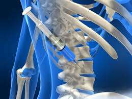
Chronic Obstructive Pulmonary Disease (COPD) encompasses progressive inflammatory conditions affecting airflow and quality of life severely. Primarily caused by long-term exposure to harmful pollutants such as tobacco smoke, it necessitates comprehensive management strategies tailored individually based upon specific patient circumstances encountered over time reflecting pathological changes occurred due airway narrowing & destruction associated chronic inflammation pathways characterizing disease progression through stages varying from mild-moderate-severe levels each requiring distinct attentiveness towards symptomatology monitoring adherence therapeutic regimens defined collaboratively specialists involved transitioning care settings smoothly between outpatient & inpatient realms accordingly maintaining proactive engagement promotes overall health sustainability amidst fluctuating challenges posed daily life symptomatic presentations experienced!
- Key indicators evident phenomenologically often include persistent cough wheezing fatigue exacerbated physical activities directly affecting functional capacities
- Compounding diagnostic efforts amalgamate spirometry essential data acquisition reflecting airflow limitations alongside pertinent imaging revealing structural insights toward chronic disease progression tracking evolution.
- Evidence-based practices suggest pharmacological approaches diversifying inhalational therapies facilitating symptomatic relief systematically managing underlying inflammatory pathophysiology contributing obstructive patterns yielding advancements therapeutic compliance facilitation empowering autonomy alongside education directives promoting self-management initiatives successfully navigating quotidian routines amidst complexities evolving components reflective personal journey traversing realms seeking optimal pulmonary health outcomes!
Managing COPD necessitates collaborative efforts facilitated effectively among diverse healthcare teams determinedly committed enhancing individual potential sustainably benefiting lives coping living coexisting demanding realities imposed constraints characterized enduring chronic illness trajectories pursued actively !
VIDEO-ASSISTED THORACOSCOPIC SURGERY (VATS)
Video-assisted thoracoscopic surgery (VATS) represents a revolutionary technique that enables surgeons to perform minimally invasive surgeries within the chest cavity. This method is particularly beneficial when treating conditions such as lung tumors, emphysema, and other thoracic pathologies requiring surgical intervention without large incisions.
- Candidates often present with symptoms related to chronic respiratory conditions requiring thorough surgical assessment and intervention strategies tailored accordingly.
- Prior diagnoses usually arise through comprehensive imaging techniques alongside physical evaluations that highlight structural issues within the lungs or pleurae needing correction surgically.
- This technique allows for quicker recovery times compared to traditional open surgeries while providing surgeons enhanced visual access through small ports alongside video equipment aiding precision operations during interventions
The VATS approach fosters earlier hospital discharge possibilities while significantly improving post-operative pain management outcomes, thereby promoting faster rehabilitation for patients following procedures related directly impacting pulmonary health!
ENDOBRONCHIAL ULTRASOUND (EBUS)

Endobronchial ultrasound (EBUS) is an advanced diagnostic method combining endoscopy with ultrasound technology. This innovative approach allows healthcare providers to visualize structures within and adjacent to the airways seamlessly. It significantly aids in staging lung cancer and evaluating lymph nodes for malignancy while minimizing invasiveness for patients.
- Symptoms prompting EBUS typically involve persistent cough or abnormal imaging findings suggesting possible lymph node involvement near bronchial structures.
- The diagnostic process generally starts with bronchoscopic visualization followed by ultrasound-guided sampling techniques to retrieve lymph node specimens accurately.
- Treatment outcomes assist in planning further procedures such as surgery or chemotherapy based on histological findings from biopsies obtained during EBUS examination.
By incorporating ultrasound technology into standard bronchoscopic practices, EBUS offers improved accuracy while ensuring patient comfort throughout evaluation processes.
PLEURAL DRAINAGE

Pleural drainage is a medical intervention used to remove excess fluid or air from the pleural space surrounding the lungs. Conditions such as pleural effusion and pneumothorax necessitate this treatment to relieve symptoms and improve lung function. The procedure can be performed in an emergency setting or electively based on clinical findings.
- Pleura-related symptoms might include shortness of breath, chest pain, and cough due to pressure impact on healthy lung tissues.
- Disease diagnosis typically requires imaging tests like chest X-rays and ultrasound before proceeding with drainage techniques.
- Treatment management involves either inserting a catheter for continuous drainage or performing thoracentesis for immediate relief of symptoms.
- The experience of draining access varies; some patients require hospitalization while others can return home shortly after completion of therapy.
This treatment significantly enhances patient comfort by alleviating respiratory distress caused by pleural accumulation of fluids or air.
LUNG BIOPSY

A lung biopsy is a critical diagnostic procedure aimed at obtaining tissue samples from lung tissue. It plays a key role in identifying various lung diseases, including infections and malignancies. This procedure can be performed using different methods such as needle biopsy or surgical biopsy depending on the lesion's location.
- Typical indications for a biopsy include suspicious nodules seen on imaging studies along with unexplained respiratory symptoms.
- The process often begins with imaging guidance—CT scans are commonly utilized for precise targeting of tissue samples.
- Treatment may involve further analysis of the obtained sample to determine if cancerous cells are present and what type they are.
- Patients usually go home the same day after observation unless complications arise.
This procedure provides essential insights necessary for tailoring an effective treatment strategy in managing lung diseases.
BRONCHOSCOPY

Bronchoscopy is a minimally invasive procedure utilized to examine the airways and lungs. This technique provides valuable information about potential lung diseases, infections, or abnormalities. During bronchoscopy, a thin tube with a camera is inserted through the nose or mouth into the lungs.
- Common symptoms that may lead to bronchoscopy include persistent cough, wheezing, or unexplained lung issues.
- The diagnosis often involves imaging tests like X-rays or CT scans prior to the procedure.
- Treatment options can range from obtaining tissue samples for biopsy to removing foreign objects obstructing the airways.
- Patients can expect some discomfort during the procedure, but sedation is typically provided.
- Post-procedure monitoring is necessary to ensure there are no complications like bleeding or infection.
Bronchoscopy is an essential tool for diagnosing and managing various pulmonary conditions effectively.