
Dr Vaidhya Raj Sh
About Me
Dr Vaidhya Raj Sh
MBBS
Cardiologist
10 Years Experience
Biography
My Clinic
OPD Hours
| Monday | Morning | 10:00 AM | - | 02:00 PM |
| Evening | 06:00 PM | - | 09:00 PM | |
| Tuesday | Morning | 10:00 AM | - | 02:00 PM |
| Evening | 06:00 PM | - | 09:00 PM | |
| Wednesday | Morning | 10:00 AM | - | 02:00 PM |
| Evening | 06:00 PM | - | 09:00 PM | |
| Thursday | Morning | 10:00 AM | - | 02:00 PM |
| Evening | 06:00 PM | - | 09:00 PM | |
| Friday | Morning | 10:00 AM | - | 02:00 PM |
| Evening | 06:00 PM | - | 09:00 PM | |
| Saturday | Morning | 10:00 AM | - | 02:00 PM |
| Evening | 06:00 PM | - | 09:00 PM | |
| Sunday | Morning | 10:00 AM | - | 02:00 PM |
| Evening | 06:00 PM | - | 09:00 PM |
Treatments
Specialised care and treatments offered
IMPLANTABLE LOOP RECORDER

An implantable loop recorder (ILR) is a small device placed under the skin that continuously monitors heart rhythms over extended periods. It is particularly useful when arrhythmias are infrequent but cause serious symptoms such as fainting spells unexplained by other tests.
- The ILR records electrical activity automatically triggered by abnormal patterns or can be activated manually during symptoms recorded in real-time for physician analysis later.
- The procedure involves minor local anesthesia inserting through a tiny incision usually done in an outpatient setting.
- This device helps pinpoint causes of transient neurological complaints possibly linked to cardiac origin.
- Treatment plans may then include medication adjustments or scheduling definitive corrective procedures triggered by ILR findings.
The ILR offers patients reassurance through comprehensive long-term rhythm surveillance not feasible via short-term external monitors while maintaining convenience and safety standards required for cardiac diagnostics today.
STRESS ECHOCARDIOGRAPHY
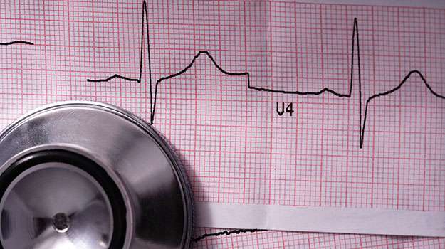
This test combines exercise or pharmacologic stress with echocardiography imaging to evaluate how well the heart functions under increased workload conditions. It helps unmask coronary artery disease not evident at rest scans alone.
- The reason it’s done: Patients complaining of exertional chest pain or shortness of breath undergo stress echo to assess inducible ischemia risk factors accurately.
HOLTER MONITORING
A Holter monitor is a portable device worn over 24-48 hours that records continuous electrocardiographic data. It enables detection of intermittent cardiac arrhythmias which might be missed during brief clinic visits.
- Syndrome indications: Patients experiencing frequent palpitations, syncope episodes, or unexplained chest discomfort benefit from this extended monitoring strategy.
- The testing mechanism: Electrodes attached on the chest connect to a compact recorder capturing heart electrical activity throughout daily routine activities including sleep cycles.
- Treatment linkage: Data guides physicians in prescribing antiarrhythmic drugs or deciding on further interventions like pacemaker implantation depending on findings severity and frequency.
Wearing this device enhances diagnosis accuracy for transient rhythm disturbances enabling tailored treatment plans based on comprehensive rhythm information collected ambulatory fashion minimally impacting lifestyle during testing period.
ECHOCARDIOGRAM EVALUATION
An echocardiogram uses ultrasound waves to produce real-time images of the heart's structure and function. It provides critical insights into diagnosing various cardiac conditions without any invasive measures.
- Pain points patients notice: Symptoms like unexplained fatigue, swelling, or irregular pulse often prompt this test for further assessment.
- The diagnostic method: The transducer device sends sound waves that reflect back from heart tissues creating detailed visuals on a monitor.
- The procedure experience: Non-invasive, painless, generally lasting 30-60 minutes performed in an outpatient setting with no radiation exposure involved.
- Disease detection capabilities: Heart valve abnormalities, chamber size changes, wall motion defects, pericardial effusions are among many conditions revealed through echo imaging.
This safe diagnostic tool plays an indispensable role for cardiologists evaluating suspected heart disease progression or response after therapies.
PERCUTANEOUS CORONARY INTERVENTION (PCI)
Percutaneous coronary intervention (PCI) is a non-surgical method used to open narrowed coronary arteries in people experiencing chest pain or heart attacks. This approach helps restore adequate blood flow to the heart muscle.
- Symptoms and causes: Blockages result from plaque buildup leading to angina or myocardial infarction symptoms like chest discomfort and breathlessness.
- Diagnostic tools: Angiography is performed by injecting contrast dye viewed under X-ray to locate arterial obstructions.
- Treatment process: A balloon catheter inflates at the blockage site followed by stent placement to keep vessels open permanently.
- Aftercare considerations: Antiplatelet therapy is essential post-PCI along with lifestyle modifications for long-term benefits.
This procedure offers a quicker recovery compared to bypass surgery while effectively alleviating ischemic symptoms and improving cardiac function.
ATRIAL FIBRILLATION ABLATION
Atrial fibrillation ablation is a minimally invasive procedure aimed at correcting abnormal heart rhythms caused by atrial fibrillation (AFib). It targets the irregular electrical signals that disrupt the heart’s natural pacing, restoring a normal heartbeat.
- Symptoms and causes: AFib may cause palpitations, shortness of breath, fatigue, or dizziness. It is often linked to high blood pressure or heart disease.
- Diagnosis: Electrocardiograms (ECG) and Holter monitoring help detect irregular heartbeats confirming AFib.
- Treatment approach: Ablation involves using catheters with energy sources such as radiofrequency to destroy problematic tissue causing erratic impulses.
- Management post-procedure: Patients usually require anticoagulants and follow-up rhythm monitoring to prevent complications.
- Expectations: Recovery is typically quick with minimal pain; success rates are favorable though repeat procedures may sometimes be necessary.
This intervention improves quality of life by reducing symptoms and lowering stroke risk associated with atrial fibrillation when medications alone are insufficient.