
Dr Shailendra Dix
About Me
Dr Shailendra Dix
MBBS
Cardiologist
10 Years Experience
Biography
My Clinic
OPD Hours
| Monday | Morning | 10:00 AM | - | 02:00 PM |
| Evening | 06:00 PM | - | 09:00 PM | |
| Tuesday | Morning | 10:00 AM | - | 02:00 PM |
| Evening | 06:00 PM | - | 09:00 PM | |
| Wednesday | Morning | 10:00 AM | - | 02:00 PM |
| Evening | 06:00 PM | - | 09:00 PM | |
| Thursday | Morning | 10:00 AM | - | 02:00 PM |
| Evening | 06:00 PM | - | 09:00 PM | |
| Friday | Morning | 10:00 AM | - | 02:00 PM |
| Evening | 06:00 PM | - | 09:00 PM | |
| Saturday | Morning | 10:00 AM | - | 02:00 PM |
| Evening | 06:00 PM | - | 09:00 PM | |
| Sunday | Morning | 10:00 AM | - | 02:00 PM |
| Evening | 06:00 PM | - | 09:00 PM |
Treatments
Specialised care and treatments offered
CARDIAC BIOMARKER TESTING

This blood test measures specific proteins released into circulation when heart muscle is damaged. It plays a key role in diagnosing myocardial infarction (heart attack) promptly upon symptom presentation.
CORONARY ANGIOGRAPHY
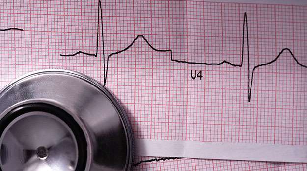
This diagnostic procedure visualizes blood flow within coronary arteries using contrast dye injected through a catheter. It identifies blockages causing chest pain or other ischemic symptoms directly within coronary vessels.
- Symptoms/causes: Recommended when there’s suspicion of coronary artery disease presenting as angina or after abnormal non-invasive tests suggest blockage risk.
- Diagnosis:The catheter is threaded through an artery (usually wrist or groin) under local anesthesia; X-ray images captured show arterial patency precisely.
- Treatment / management : Besides diagnosis , angioplasty with stent placement can be performed immediately if significant obstruction is found .
- What to expect : The procedure usually lasts 30 - 60 minutes ; patients may feel minimal discomfort during catheter insertion but rest afterward is required .
This method remains one of the gold standards for evaluating coronary artery disease allowing prompt therapeutic decisions .
HOLTER MONITORING
The Holter Monitor is a portable device worn for 24-48 hours to continuously record your heart's electrical activity during normal daily routines. It detects intermittent arrhythmias that may be missed by standard ECGs.
- Symptoms/causes: Indicated if experiencing irregular heartbeats, fainting episodes, dizziness or palpitations occurring sporadically over days.
- Diagnosis: Small electrodes attached to the chest connect to the monitor worn around the waist; it logs data automatically throughout the day.
- Treatment/management: Data from the Holter helps tailor treatments such as medications for arrhythmia control or recommendations for further electrophysiological studies.
- What to expect: You can maintain usual activities but should avoid water contact; afterwards results are reviewed by specialists for accurate interpretation.
This continuous monitoring method provides critical insights into transient cardiac irregularities enabling timely intervention and improved outcomes.
STRESS TESTING (EXERCISE STRESS TEST)
A Stress Test evaluates how well your heart functions during physical activity. It helps identify blockages in coronary arteries that might not be apparent at rest.
- Symptoms/causes: Useful for patients with chest discomfort on exertion, unexplained fatigue during activity, or risk factors like diabetes and hypertension.
- Diagnosis: Patient walks on a treadmill or pedals a stationary bike while their ECG and blood pressure are continuously monitored.
- Treatment/management: Abnormal findings can lead to recommendations such as medication changes, further imaging tests, or interventions like angioplasty.
- What to expect: The duration varies but generally lasts about 30-45 minutes; you will be closely observed by trained staff throughout the test.
This non-invasive procedure provides valuable information about exercise tolerance and cardiovascular health under stress conditions safely.
ECHOCARDIOGRAPHY
Echocardiography uses ultrasound waves to create moving images of the heart. This imaging technique plays a crucial role in evaluating heart structure and function in real time.
- Symptoms/causes: Recommended for patients with suspected valve disease, heart failure symptoms, murmurs, or unexplained breathlessness.
- Diagnosis: A transducer sends sound waves through the chest wall which bounce back from heart structures to create detailed visuals.
- Treatment/management: Results provide insights for managing conditions such as valve repair needs, cardiomyopathies, or monitoring treatment effectiveness.
- What to expect: The procedure is safe and painless; typically takes 30-60 minutes and may require lying on your side during the scan.
This approach offers comprehensive insight into cardiac anatomy without radiation exposure aiding accurate diagnosis and personalized treatment plans.
ELECTROCARDIOGRAM (ECG)
An Electrocardiogram, commonly known as ECG or EKG, is a simple and painless test that records the electrical activity of your heart over time. It's essential for assessing heart rhythm and detecting various cardiac issues.
- Symptoms/causes: Often used when patients experience chest pain, palpitations, dizziness, or shortness of breath to detect arrhythmias or ischemia.
- Diagnosis: Electrodes placed on the skin capture electrical impulses which produce waveforms on paper or screen for analysis.
- Treatment/management: Helps guide further management like medication adjustment, lifestyle changes, or advanced testing if abnormalities are found.
- What to expect: The procedure is quick, typically under 10 minutes with no discomfort; results are immediate and non-invasive.
This test serves as a foundational tool in cardiac care by providing critical information about heart health quickly and safely.