
Dr Reena Jain
About Me
Dr Reena Jain
MS
Orthopaedic Surgeons
10 Years Experience
Biography
My Clinic
OPD Hours
| Monday | Morning | 10:00 AM | - | 02:00 PM |
| Evening | 06:00 PM | - | 09:00 PM | |
| Tuesday | Morning | 10:00 AM | - | 02:00 PM |
| Evening | 06:00 PM | - | 09:00 PM | |
| Wednesday | Morning | 10:00 AM | - | 02:00 PM |
| Evening | 06:00 PM | - | 09:00 PM | |
| Thursday | Morning | 10:00 AM | - | 02:00 PM |
| Evening | 06:00 PM | - | 09:00 PM | |
| Friday | Morning | 10:00 AM | - | 02:00 PM |
| Evening | 06:00 PM | - | 09:00 PM | |
| Saturday | Morning | 10:00 AM | - | 02:00 PM |
| Evening | 06:00 PM | - | 09:00 PM | |
| Sunday | Morning | 10:00 AM | - | 02:00 PM |
| Evening | 06:00 PM | - | 09:00 PM |
Treatments
Specialised care and treatments offered
BONE DENSITY TEST
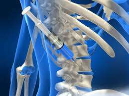
A bone density test measures mineral content within bones using specialized equipment like dual-energy X-ray absorptiometry (DXA). This assessment helps identify osteoporosis risk before fractures occur.
- Symptoms/causes: Often associated with aging or hormonal changes, individuals may be recommended this test if they exhibit signs of weakened bone structure such as frequent fractures without major trauma.
- Diagnosis: The test is simple; patients lie down while scanners pass over their body measuring bone density at specific sites like hips or spine regions.
- Treatment options: After analysis revealed low-density levels indicating osteoporosis risk factors intervention plans could involve lifestyle modifications along with medications aimed at enhancing bone strength over time.
- What to expect: The procedure lasts about 10-30 minutes; results are typically shared soon after providing insights necessary for proactive management strategies going forward.
Regular monitoring via such tests contributes immensely towards early detection hence fortifying preventive measures against osteoporosis related complications later downline!
ARTHROSCOPY

An arthroscopy is a minimally invasive surgical procedure that allows doctors to examine and treat joint disorders through small incisions. It’s particularly beneficial for diagnosing issues not visible via other forms of imaging technology.
- Symptoms/causes: Patients often undergo this procedure due to unexplained joint pain or mechanical symptoms such as locking or catching during movement.
- Diagnosis: During arthroscopy, a small camera (arthroscope) is inserted into the joint through tiny incisions providing real-time visualization on a monitor.
- Treatment options: Not only can this technique provide diagnosis but also allows for immediate treatment measures ranging from debridement (cleaning) of damaged tissue to ligament repairs.
- What to expect: Recovery times can vary depending on the complexity but many patients experience minimal discomfort post-surgery and follow-up care usually proceeds effectively after just a few days.
Arthroscopy enhances comprehension regarding intricate joint problems while facilitating targeted therapeutic interventions promptly.
ULTRASOUND IMAGING

Ultrasound imaging provides real-time visualization of musculoskeletal structures using sound waves. It’s particularly useful for assessing joint and soft tissue conditions without exposure to radiation.
- Symptoms/causes: Pain or swelling around joints may prompt healthcare professionals to recommend an ultrasound exam for accurate evaluation.
- Diagnosis: By placing a transducer over the area of interest, doctors can view live images highlighting any abnormalities or fluid buildup.
- Treatment/management options: Results can lead to treatments ranging from physical therapy recommendations to potential minimally invasive procedures if necessary.
- What to expect: The examination process is quick—typically completed within 15-30 minutes—and there is no downtime afterwards as it’s non-invasive。
This technique offers direct insight into musculoskeletal conditions, aiding physicians in making informed decisions regarding patient care
CT SCAN

A CT scan combines multiple X-ray images to produce cross-sectional views of bones and organs. It is widely utilized in orthopaedic practice for its ability to reveal complex structures more effectively than standard X-rays.
- Symptoms/causes: Individuals may require a CT scan when experiencing significant trauma or persistent pain that remains undiagnosed through basic imaging techniques.
- Diagnosis: During the examination, patients lie still while the scanner rotates around them, capturing numerous images quickly.
- Treatment options: Based on findings, rehabilitation or corrective surgery could be recommended based on detailed analysis of injuries or abnormalities identified.
- What to expect: The procedure is painless and usually takes only a few minutes; radiologists will analyze the images for interpretation shortly thereafter.
This advanced imaging modality plays a vital role in comprehensive orthopaedic assessments and planning effective treatments.
MRI SCAN

An MRI scan serves as a powerful diagnostic method in orthopaedics, allowing for detailed imaging of soft tissues like muscles, ligaments, and cartilage. It plays an integral role in assessing joint conditions.
- Symptoms/causes: Patients often experience unexplained pain or restricted movement that warrants further investigation via MRI.
- Diagnosis: This non-invasive technique utilizes strong magnets and radio waves to create comprehensive images of the body’s internal structures.
- Treatment/management options: Depending on results, patients might be advised on physiotherapy or surgical procedures if necessary.
- What to expect: The scanning process lasts about 30-60 minutes; however, no discomfort is typically felt by patients during the session.
The clarity provided by MRI can significantly enhance treatment accuracy and outcomes for various conditions.
X-RAY IMAGING

X-ray imaging is a crucial diagnostic tool used to visualize bones and joints. It helps in identifying various conditions based on the density of bone structures and is commonly employed in orthopaedic evaluations.
- Symptoms/causes: Patients may present with pain, swelling, or trauma that necessitates an X-ray for clear diagnosis.
- Diagnosis: The procedure involves positioning the patient and directing low-dose radiation to capture images of the skeletal structure.
- Treatment options: Depending on findings, treatment may range from medication for inflammation to surgical interventions.
- What to expect: The entire process is quick, often completed within minutes, with results typically ready for review shortly thereafter.
X-rays are essential in providing a clear picture of bone integrity, assisting in effective patient management.