
Dr R L Meena
About Me
Dr R L Meena
MS
Orthopaedic Surgeons
10 Years Experience
Biography
My Clinic
OPD Hours
| Monday | Morning | 10:00 AM | - | 02:00 PM |
| Evening | 06:00 PM | - | 09:00 PM | |
| Tuesday | Morning | 10:00 AM | - | 02:00 PM |
| Evening | 06:00 PM | - | 09:00 PM | |
| Wednesday | Morning | 10:00 AM | - | 02:00 PM |
| Evening | 06:00 PM | - | 09:00 PM | |
| Thursday | Morning | 10:00 AM | - | 02:00 PM |
| Evening | 06:00 PM | - | 09:00 PM | |
| Friday | Morning | 10:00 AM | - | 02:00 PM |
| Evening | 06:00 PM | - | 09:00 PM | |
| Saturday | Morning | 10:00 AM | - | 02:00 PM |
| Evening | 06:00 PM | - | 09:00 PM | |
| Sunday | Morning | 10:00 AM | - | 02:00 PM |
| Evening | 06:00 PM | - | 09:00 PM |
Treatments
Specialised care and treatments offered
ULTRASOUND IMAGING
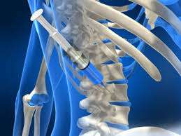
Ultrasound imaging uses sound waves to create real-time images of soft tissues around joints. It is especially valuable in guiding procedures like injections or aspirations directly into affected areas without radiation exposure.
- Pain connected with inflammation or fluid accumulation can trigger an ultrasound assessment for precise evaluation of soft tissue conditions affecting the joints.
- The technician applies gel on the skin while moving a transducer over the area of interest; sound waves produce detailed pictures instantly
- Conditions diagnosed via ultrasound include tendonitis, bursitis, tears in muscles or ligaments caused by overuse.
- Based on ultrasound findings, doctors can offer targeted treatments such as corticosteroid injections directly into inflamed areas
This non-invasive approach enhances diagnosis accuracy while minimizing discomfort during evaluations providing maximum benefits for patient management
BONE DENSITY TEST

A bone density test measures the amount of mineral content (primarily calcium) in your bones. This test is crucial for assessing bone health and diagnosing osteoporosis before significant fractures occur.
- Common indicators prompting this test include family history of osteoporosis and prior fractures after minimal trauma.
- The procedure is quick and painless; it typically involves lying on a padded table while low-dose X-rays scan your spine and hips or other areas at risk for fracture.
- Low results indicate weaker bones that may require preventive measures such as lifestyle changes or medication.
- Managing osteoporosis often involves supplements, dietary alterations, exercise programs aimed at strengthening bones, and sometimes medication.
Early detection through bone density testing promotes proactive management strategies ensuring long-term skeletal health
CT SCAN

A CT scan combines X-ray technology with computer processing to produce cross-sectional images of bones and joints. This advanced imaging method offers more detailed insight into complex fractures or joint diseases than regular X-rays alone.
- Severe trauma cases often lead physicians to recommend a CT scan due to its ability to display intricate skeletal details.
- This process entails using a series of X-ray images taken from various angles, which are then reconstructed by a computer into detailed slices of the body structure being examined.
- CT scans play a critical role in diagnosing conditions like bone tumors or assessing post-surgical anatomy after joint replacements.
- Management strategies depend on CT findings and may involve surgical interventions when necessary for optimal outcomes.
The precision offered by CT scans significantly aids in effective treatment planning for orthopedic conditions.
MRI SCAN

An MRI scan provides detailed images of soft tissues, cartilage, and ligaments around joints. It is particularly useful in diagnosing sports-related injuries without exposing patients to radiation.
- Patients with unexplained pain or swelling will often undergo an MRI as part of their diagnostic workup.
- The scan utilizes powerful magnets and radio waves to create comprehensive images of internal structures.
- MRI findings can reveal tears in ligaments or cartilage damage that may not be visible on X-rays.
- Treatment options based on MRI results range from conservative care such as physiotherapy to more invasive surgical procedures if required.
This advanced imaging technique is crucial for understanding complex musculoskeletal conditions effectively.
X-RAY IMAGING

X-ray imaging is an essential diagnostic tool in orthopaedics that helps in visualizing bone structures and detecting fractures or abnormalities. It’s a quick and non-invasive method to assess injuries.
- Painful symptoms such as swelling or inability to move a limb may prompt an X-ray examination.
- The process involves positioning the patient correctly to capture clear images of the specific area being examined.
- X-rays help identify conditions like fractures, arthritis, or tumors in the bones.
- The findings guide treatment decisions ranging from conservative management with medication to surgical interventions if necessary.
X-rays are vital for formulating effective treatment plans tailored to individual patient needs.
ARTHROSCOPY

Arthroscopy is a minimally invasive surgical procedure that allows orthopaedic surgeons to visualize, diagnose, and treat joint issues. It's commonly used for knee, shoulder, and ankle surgeries.
- Symptoms of conditions that may require arthroscopy include persistent joint pain, swelling, and restricted mobility.
- The procedure typically involves inserting a small camera into the joint through small incisions, allowing for accurate diagnosis.
- Treatment options may involve removing damaged tissue or repairing ligaments using specialized instruments.
- Recovery often includes physical therapy to restore function and strength in the affected area.
This technique offers quicker recovery times compared to traditional surgery, making it a preferred choice for many patients.