
Dr Priyadarshani
About Me
Dr Priyadarshani
MBBS
Ophthalmologist
10 Years Experience
Biography
My Clinic
OPD Hours
| Monday | Morning | 10:00 AM | - | 02:00 PM |
| Evening | 06:00 PM | - | 09:00 PM | |
| Tuesday | Morning | 10:00 AM | - | 02:00 PM |
| Evening | 06:00 PM | - | 09:00 PM | |
| Wednesday | Morning | 10:00 AM | - | 02:00 PM |
| Evening | 06:00 PM | - | 09:00 PM | |
| Thursday | Morning | 10:00 AM | - | 02:00 PM |
| Evening | 06:00 PM | - | 09:00 PM | |
| Friday | Morning | 10:00 AM | - | 02:00 PM |
| Evening | 06:00 PM | - | 09:00 PM | |
| Saturday | Morning | 10:00 AM | - | 02:00 PM |
| Evening | 06:00 PM | - | 09:00 PM | |
| Sunday | Morning | 10:00 AM | - | 02:00 PM |
| Evening | 06:00 PM | - | 09:00 PM |
Treatments
Specialised care and treatments offered
RETINAL LASER PHOTOCOAGULATION
This important laser therapy treats various retinal vascular disorders by sealing leaking blood vessels and creating scar tissue that reinforces weakened areas inside the retina helping prevent further deterioration of sight over time.
CORNEAL TRANSPLANTATION (KERATOPLASTY)

This surgery replaces a damaged or diseased cornea with healthy donor tissue. It restores clarity when scarring or other conditions severely impair corneal transparency affecting vision quality dramatically.
VITRECTOMY

A vitrectomy involves surgically removing the vitreous gel from inside the eye. This operation treats various retinal disorders by providing access for repair and clearing blood or scar tissue obstructing vision.
- Main Indications: Conditions like retinal detachment, diabetic retinopathy complications, or vitreous hemorrhage may require vitrectomy intervention.
- Diagnostic Assessment: Retinal imaging including OCT scans aids precise diagnosis and planning before surgery.
- Surgical Process Overview: Small incisions allow instruments into the eye to remove vitreous gel; damaged retina can then be repaired or membranes peeled away carefully.
- Aim of Treatment:
The procedure demands specialized expertise but can be crucial in salvaging sight in complex retinal diseases.
GLAUCOMA DRAINAGE DEVICE IMPLANTATION

This surgical intervention is designed to manage glaucoma cases where traditional medications or laser treatments have failed to adequately control eye pressure. It facilitates fluid drainage from the eye to prevent optic nerve damage.
- Causative Factors & Symptoms: Often due to impaired drainage channels causing elevated intraocular pressure; symptoms can include peripheral vision loss.
- Evaluation Techniques: Measurement of intraocular pressure and optic nerve assessment guide diagnosis and treatment planning.
- Surgical Approach: Implantation of a tiny tube connected to a reservoir plate that redirects aqueous humor outflow to lower eye pressure effectively.
- Treatment Expectations: Postoperative monitoring ensures device function; adjunctive medications may still be necessary initially.
This procedure offers hope for preserving remaining vision in advanced or refractory glaucoma cases through controlled pressure reduction.
LASER ASSISTED IN SITU KERATOMILEUSIS (LASIK)
LASIK is a popular refractive surgery aimed at correcting refractive errors such as myopia, hyperopia, and astigmatism. It helps reduce dependence on corrective lenses.
- Identifying Candidates: Suitable for individuals with stable prescriptions and healthy corneas.
- Diagnosis: Detailed corneal mapping and ocular measurements are performed to design the laser correction plan.
- Surgical Process: A flap in the cornea is created, reshaped using an excimer laser, then repositioned without stitches.
- Recovery: Patients often notice clearer vision within 24 hours; minor discomfort may be experienced initially.
- Long-Term Results: Many achieve 20/20 vision or better with lasting effects, though some may require enhancements.
The precision of LASIK offers rapid visual improvement with minimal downtime for suitable candidates.
CATARACT SURGERY
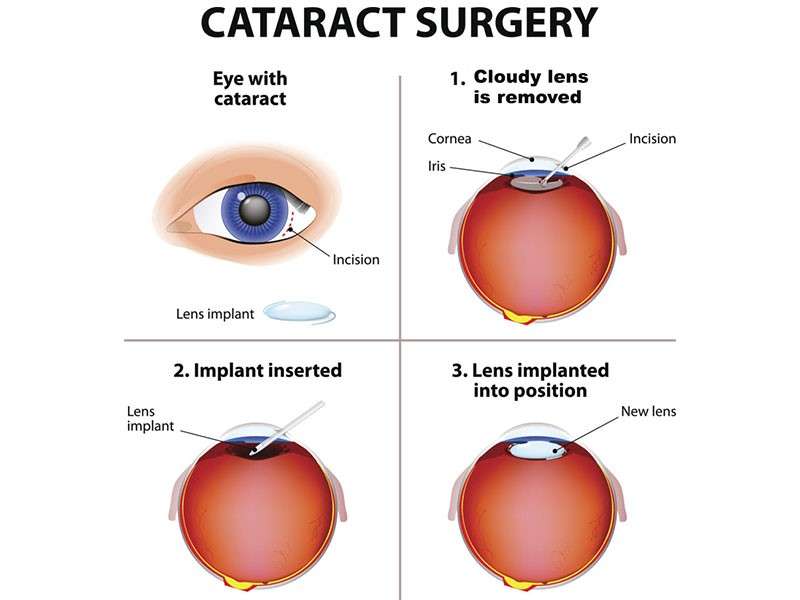
Cataracts cause clouding of the eye’s natural lens and can significantly impair vision. Surgery is often needed to restore sight when cataracts progress beyond mild impairment.
- Symptoms and Causes: Gradual blurring or dimming of vision, glare sensitivity, and difficulty seeing at night; aging is the primary cause.
- Diagnosis: Comprehensive eye examination including slit-lamp evaluation and visual acuity testing.
- Treatment Options: The cloudy lens is removed and typically replaced with a clear, artificial intraocular lens (IOL).
- Procedural Details: Usually an outpatient procedure using local anesthesia with minimal discomfort.
- Expected Outcomes: Most patients experience improved vision within days to weeks following surgery.
This treatment remains one of the safest and most effective means of restoring vision affected by cataracts, offering patients a better quality of life.