
Dr Prashant Rast
About Me
Dr Prashant Rast
MBBS
Cardiologist
10 Years Experience
Biography
My Clinic
OPD Hours
| Monday | Morning | 10:00 AM | - | 02:00 PM |
| Evening | 06:00 PM | - | 09:00 PM | |
| Tuesday | Morning | 10:00 AM | - | 02:00 PM |
| Evening | 06:00 PM | - | 09:00 PM | |
| Wednesday | Morning | 10:00 AM | - | 02:00 PM |
| Evening | 06:00 PM | - | 09:00 PM | |
| Thursday | Morning | 10:00 AM | - | 02:00 PM |
| Evening | 06:00 PM | - | 09:00 PM | |
| Friday | Morning | 10:00 AM | - | 02:00 PM |
| Evening | 06:00 PM | - | 09:00 PM | |
| Saturday | Morning | 10:00 AM | - | 02:00 PM |
| Evening | 06:00 PM | - | 09:00 PM | |
| Sunday | Morning | 10:00 AM | - | 02:00 PM |
| Evening | 06:00 PM | - | 09:00 PM |
Treatments
Specialised care and treatments offered
BLOOD PRESSURE MONITORING (AMBULATORY)

CORONARY ANGIOGRAPHY
This procedure involves imaging the coronary arteries using contrast dye injected through a catheter to visualize blockages or abnormalities under X-ray guidance. It remains the gold standard for diagnosing coronary artery disease (CAD).
- Pertinent indications:Persistent chest pain suggestive of angina unresponsive to medications or inconclusive non-invasive tests warrant angiography evaluation.
- The procedure outline:A catheter is advanced through an artery typically from the wrist or groin into coronary vessels; contrast dye illuminates arteries enabling precise mapping of narrowing spots or occlusion sites.
HOLTER MONITORING
A Holter monitor is a portable device that continuously records the electrical activity of your heart over 24 to 48 hours. This extended monitoring helps detect irregular heart rhythms that may not appear during a brief doctor’s visit or standard ECG recording.
- When it's used: Prescribed for patients experiencing intermittent palpitations, dizziness, syncope attacks, or unexplained fainting spells.
- The device setup: Small electrodes attach to the chest connected via wires to a compact recorder worn throughout normal daily activities including sleep.
- Data analysis:The recorded information is reviewed by specialists to identify arrhythmias like atrial fibrillation or ventricular ectopy occurring sporadically but significantly affecting health.
- No restrictions needed:You can carry out routine tasks ensuring an accurate representation of your heartbeat in everyday life scenarios.
This technique improves diagnostic accuracy of transient cardiac events not captured during short-term clinical evaluations.
STRESS TEST (EXERCISE TREADMILL TEST)
A stress test evaluates how well the heart performs under physical exertion. It helps identify coronary artery disease and other functional cardiac issues by monitoring symptoms and heart responses during controlled exercise.
- Main objective: Detect reduced blood flow to the heart muscle during activity when symptoms might be more evident.
- Process: The patient walks on a treadmill or pedals a stationary bike while the ECG and blood pressure are continuously monitored.
- Indicators observed: Includes chest discomfort, changes in ECG patterns, abnormal blood pressure response, or arrhythmias triggered by exercise.
- Safety considerations: Monitored closely by medical staff to promptly address any adverse reactions or complications.
This dynamic assessment offers critical insight beyond resting tests by revealing exercise-induced cardiac problems that might otherwise go undetected at rest.
ELECTROCARDIOGRAM (ECG)
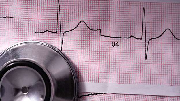
An electrocardiogram (ECG) records the electrical activity of the heart over time. It is one of the simplest and most widely used tests for evaluating heart rhythm and detecting abnormalities that may indicate underlying cardiac issues.
- Symptoms assessed: Includes chest pain, palpitations, dizziness, or unexplained shortness of breath.
- How it works: Electrodes placed on the skin measure electrical impulses produced during each heartbeat.
- Identification capabilities: Can detect arrhythmias, ischemia, previous heart attacks, and electrolyte imbalances.
- Procedure details: The test lasts about 5-10 minutes and is completely painless with no risks involved.
An ECG serves as a frontline screening method to quickly evaluate cardiac status and often guides further diagnostic testing or treatment plans.
ECHOCARDIOGRAM
An echocardiogram is a crucial non-invasive imaging test that uses ultrasound waves to create moving pictures of the heart. It allows physicians to assess heart structure and function in real-time, providing detailed information essential for diagnosing various cardiac conditions.
- Purpose: Evaluates heart chambers, valves, and overall pumping ability to detect abnormalities.
- Procedure: A transducer placed on the chest sends sound waves that bounce off heart structures, creating images on a monitor.
- Conditions diagnosed: Includes valve disorders, cardiomyopathies, pericardial effusion, and congenital defects.
- Duration & Comfort: Typically takes 30-60 minutes; it is painless and requires no special preparation.
- Follow-up: Results guide treatment decisions such as medication adjustments or need for further testing like stress echocardiography.
This test provides valuable insights into heart health without exposure to radiation or invasive procedures, making it a standard diagnostic tool in cardiology.