
Dr P K Bhatnaagar
About Me
Dr P K Bhatnaagar
MBBS
Cardiologist
10 Years Experience
Biography
My Clinic
OPD Hours
| Monday | Morning | 10:00 AM | - | 02:00 PM |
| Evening | 06:00 PM | - | 09:00 PM | |
| Tuesday | Morning | 10:00 AM | - | 02:00 PM |
| Evening | 06:00 PM | - | 09:00 PM | |
| Wednesday | Morning | 10:00 AM | - | 02:00 PM |
| Evening | 06:00 PM | - | 09:00 PM | |
| Thursday | Morning | 10:00 AM | - | 02:00 PM |
| Evening | 06:00 PM | - | 09:00 PM | |
| Friday | Morning | 10:00 AM | - | 02:00 PM |
| Evening | 06:00 PM | - | 09:00 PM | |
| Saturday | Morning | 10:00 AM | - | 02:00 PM |
| Evening | 06:00 PM | - | 09:00 PM | |
| Sunday | Morning | 10:00 AM | - | 02:00 PM |
| Evening | 06:00 PM | - | 09:00 PM |
Treatments
Specialised care and treatments offered
BLOOD PRESSURE MONITORING (AMBULATORY)

The Ambulatory Blood Pressure Monitor records blood pressure throughout daily activities over 24 hours. This provides a comprehensive profile compared to single clinic measurements which can be misleading due to fluctuations caused by stress or environment.
- Symptoms and causes: Used when there's suspicion of white-coat hypertension (elevated readings only at clinics) or masked hypertension undetected in-office but harmful long term.
- How it is diagnosed: A cuff worn on the arm automatically inflates at preset intervals collecting multiple readings stored digitally for later analysis by cardiologists.
- Treatment and management options: More accurate BP profiles enable personalized antihypertensive therapy adjustments improving control reducing risks associated with uncontrolled high blood pressure.
- What to expect: Device worn comfortably all day including sleep periods but regular movement may cause some mild discomfort from cuff inflations intermittently. Diaries noting activities alongside readings can aid interpretation.
This method enhances diagnosis accuracy for hypertension enabling better cardiovascular risk reduction strategies long-term.
CORONARY ANGIOGRAPHY
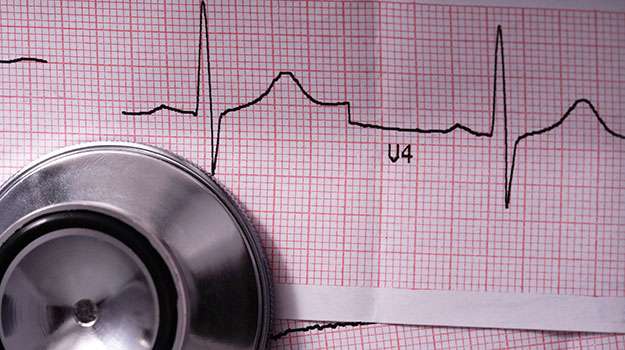
This invasive procedure visualizes coronary arteries using contrast dye injected via a catheter guided through blood vessels. It maps out blockages contributing to chest pain or acute coronary syndromes.
- Symptoms and causes: Performed when non-invasive tests suggest significant coronary artery disease causing angina or after myocardial infarction diagnosis for treatment planning.
- How it is diagnosed: X-ray fluoroscopy captures images showing areas of narrowing/blockage within coronary vessels following dye injection through catheterization typically via wrist or groin artery access points.
- Treatment and management options: Depending on findings angioplasty with stent placement may be done immediately; otherwise medications optimized based on severity assessed visually here.
- What to expect: Procedure usually takes less than an hour under local anesthesia with short recovery periods though hospitalization might extend based on interventions performed afterward.
Due its precision this technique remains gold-standard for coronary evaluation allowing timely targeted treatment decisions.
HOLTER MONITORING
A Holter Monitor tracks your heart's electrical activity continuously over 24-48 hours during normal daily routines. This ambulatory ECG helps detect intermittent arrhythmias that brief exams may miss.
- Symptoms and causes: Recommended for patients with sporadic palpitations, syncope episodes, unexplained dizziness or suspected intermittent arrhythmias.
- How it is diagnosed: Small electrodes attached to your chest connect via wires to a portable recorder worn at waist level or around the neck.
- Treatment and management options: Data analyzed later reveals abnormal rhythms guiding medication adjustments or further investigations if needed.
- What to expect: You can continue most activities but should avoid water bathing; maintain diary notes correlating symptoms with recorded events if possible.
This monitoring method provides valuable extended insight into heartbeat irregularities beyond clinical visits’ snapshots.
STRESS TESTING (TREADMILL TEST)
A Stress Test evaluates how your heart performs under controlled physical exertion. By monitoring heart rate, rhythm, blood pressure, and symptoms during exercise, it uncovers issues not evident at rest.
- Symptoms and causes: Commonly performed when patients report chest discomfort during activity or have risk factors for coronary artery disease.
- How it is diagnosed: You walk on a treadmill with gradually increasing intensity while connected to an ECG monitor; blood pressure is periodically checked.
- Treatment and management options: Abnormal responses may prompt further imaging tests or therapeutic strategies like lifestyle changes or medications.
- What to expect: The test lasts approximately 10-15 minutes but preparation includes resting ECGs before starting; cessation occurs once target heart rate or symptoms appear.
This evaluation provides crucial information on cardiovascular fitness levels and underlying ischemic conditions safely under supervision.
ECHOCARDIOGRAPHY
Echocardiography uses ultrasound waves to produce images of the heart's structure and function. It offers dynamic visualization of heart chambers, valves, and blood flow without any radiation exposure.
- Symptoms and causes: Indicated when there are signs of heart failure, murmurs, or suspected valve diseases.
- How it is diagnosed: A transducer emits sound waves that bounce off cardiac tissues to generate real-time images on screen.
- Treatment and management options: Findings help assess ventricular function, valve integrity, congenital defects, guiding medical or surgical interventions.
- What to expect: The exam typically lasts 30-60 minutes; it is painless and requires lying still while a technician moves the probe around your chest.
This imaging test is fundamental in evaluating many cardiovascular conditions effectively and safely.
ELECTROCARDIOGRAM (ECG)
An Electrocardiogram, or ECG, is a straightforward and essential diagnostic tool used in cardiology to record the electrical activity of the heart over time. It helps detect various heart conditions by measuring electrical signals as the heart beats.
- Symptoms and causes: It is frequently recommended for patients experiencing chest pain, palpitations, dizziness, or unexplained fatigue, as well as screening for arrhythmias or myocardial ischemia.
- How it is diagnosed: Electrodes placed on the skin capture electrical impulses that are graphically displayed on a monitor or printed on paper.
- Treatment and management options: Results from the ECG guide physicians in diagnosing arrhythmias, heart attacks, and other cardiac abnormalities; further testing or treatment plans are based on these findings.
- What to expect: The procedure is painless, non-invasive, takes about 5-10 minutes, and involves minimal preparation.
This test provides rapid insight into cardiac health and plays a critical role in both emergency and routine cardiac care.