Dr K P Chandra Di
About Me
Dr K P Chandra Di
MBBS
Cardiothorocic Surgeon
10 Years Experience
Biography
My Clinic
OPD Hours
| Monday | Morning | 10:00 AM | - | 02:00 PM |
| Evening | 06:00 PM | - | 09:00 PM | |
| Tuesday | Morning | 10:00 AM | - | 02:00 PM |
| Evening | 06:00 PM | - | 09:00 PM | |
| Wednesday | Morning | 10:00 AM | - | 02:00 PM |
| Evening | 06:00 PM | - | 09:00 PM | |
| Thursday | Morning | 10:00 AM | - | 02:00 PM |
| Evening | 06:00 PM | - | 09:00 PM | |
| Friday | Morning | 10:00 AM | - | 02:00 PM |
| Evening | 06:00 PM | - | 09:00 PM | |
| Saturday | Morning | 10:00 AM | - | 02:00 PM |
| Evening | 06:00 PM | - | 09:00 PM | |
| Sunday | Morning | 10:00 AM | - | 02:00 PM |
| Evening | 06:00 PM | - | 09:00 PM |
Treatments
Specialised care and treatments offered
ROBOTIC-ASSISTED MITRAL VALVE REPAIR
This cutting-edge approach enables precise mitral valve repair utilizing robotic instruments controlled by the surgeon from a console. Small ports replace large chest incisions traditionally required in open surgery.
TRANSCATHETER AORTIC VALVE REPLACEMENT (TAVR)
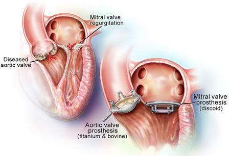
TAVR is a groundbreaking alternative for replacing a diseased aortic valve without open-heart surgery. It uses catheter-based delivery of a new valve through blood vessels.
- Disease Overview: Used primarily in severe aortic stenosis where valve narrowing restricts blood flow from the heart.
- Pre-Procedure Tests: Echocardiograms and CT scans assess valve anatomy and vascular access routes before intervention.
- The Procedure Itself: A collapsible replacement valve is threaded via femoral artery or other access points into position inside the damaged valve.
- Main Benefits: Significantly less invasive with faster recovery times suitable especially for high-risk surgical patients.
ENDOSCOPIC THORACIC SYMPATHECTOMY
This procedure is designed to treat excessive sweating (hyperhidrosis) or certain circulatory problems by interrupting nerves in the chest. It uses endoscopic tools inserted through tiny incisions.
- Conditions Treated: Commonly addresses severe palmar hyperhidrosis (excessive hand sweating) and Raynaud’s phenomenon affecting fingers.
- Assessment: Detailed clinical evaluation confirms symptom severity and suitability for surgery.
- Surgical Process: Small cameras guide nerve interruption or removal via minimal incisions under general anesthesia.
- Efficacy and Safety: Provides significant symptom relief with low complication rates compared to open procedures.
- Recovery Details: Hospital stays are brief; most resume normal activities within a week post-surgery.
This minimally invasive method effectively targets nerve pathways responsible for certain autonomic dysfunctions while ensuring rapid convalescence.
VIDEO-ASSISTED THORACOSCOPIC SURGERY (VATS) FOR LUNG BIOPSY
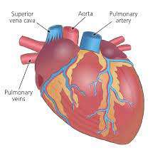
VATS is a minimally invasive procedure used to obtain lung tissue samples when diagnosing lung diseases. It provides excellent visualization of the lungs through small chest incisions.
- Indications: Used when imaging shows suspicious lung nodules or masses requiring tissue diagnosis.
- Diagnostic Tools: Preoperative CT scans help guide the biopsy location precisely.
- Surgical Technique: A thoracoscope and specialized instruments are inserted through small openings between ribs under general anesthesia.
- Advantages: Less postoperative pain, shorter recovery than traditional open lung biopsy methods.
- Follow-Up: Pathology results inform further treatment plans; patients often recover fully within days to weeks.
This approach allows safe and effective lung tissue sampling, aiding in accurate diagnosis with minimal disruption to normal anatomy.
MINIMALLY INVASIVE CORONARY ARTERY BYPASS SURGERY
Minimally invasive coronary artery bypass surgery is a less traumatic option for patients needing improved blood flow to the heart. It avoids the traditional large chest incision, promoting quicker recovery.
- Symptoms and Causes: Often recommended for patients with blocked coronary arteries causing chest pain or risk of heart attack.
- Diagnosis: Confirmed by imaging tests like coronary angiography and stress tests to locate blockages.
- Treatment Approach: Small incisions allow surgeons to reroute blood vessels around blocked arteries without stopping the heart.
- Benefits: Reduced pain, shorter hospital stay, faster return to daily activities compared to open surgery.
- Postoperative Expectations: Patients need follow-up for wound care and cardiac rehabilitation to ensure successful recovery.
This technique offers an effective way to restore blood supply while minimizing surgical trauma, making it a preferred choice for suitable candidates.
VENTRICULAR ASSIST DEVICE (VAD) IMPLANTATION
Ventricular Assist Device (VAD) implantation is a surgical procedure that involves the placement of a mechanical pump or device to assist the pumping function of a weakened or failing heart. VADs are primarily used as a bridge to heart transplantation or as destination therapy for patients who are not eligible for heart transplantation.
VAD implantation is considered for individuals with severe heart failure who have exhausted other treatment options and have a poor prognosis without intervention. A thorough evaluation is performed to assess the patient's overall health, heart function, and suitability for VAD therapy.
There are different types of VADs available, including:
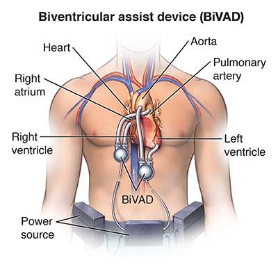
- Left Ventricular Assist Device (LVAD): This device is implanted to assist the pumping function of the left ventricle, which is the main pumping chamber of the heart.
- Right Ventricular Assist Device (RVAD): In some cases, a separate device may be implanted to support the right ventricle's function.
- Biventricular Assist Device (BiVAD): This involves the implantation of both an LVAD and an RVAD to support both ventricles.
- Total Artificial Heart: In rare cases of severe biventricular failure, a total artificial heart may be implanted to replace both ventricles temporarily until a heart transplant becomes available.
The VAD implantation procedure is typically performed under general anesthesia. The surgeon makes an incision in the chest and places the device in the appropriate position, which can be in the abdomen or the chest cavity. Tubes are connected to the heart chambers to divert blood flow through the VAD. The VAD is then connected to an external power source and controller.
Following the surgery, the patient is closely monitored in the intensive care unit (ICU) to ensure proper functioning of the VAD, stability of the patient, and to manage any immediate complications. The patient may remain in the hospital for several weeks to receive specialized care and support during the recovery phase.
Once the patient is stable, a rehabilitation program may be initiated to promote physical recovery and help the patient regain strength. The patient and their caregivers also receive education and training on VAD management, including how to maintain and troubleshoot the device, manage medications, and recognize signs of potential complications.
After leaving the hospital, the patient continues to receive regular follow-up care from a specialized VAD team. This includes monitoring VAD function, managing medications, conducting routine tests, and addressing any issues or concerns that may arise.
Ventricular Assist Devices can significantly improve the quality of life and survival for individuals with advanced heart failure. While VADs can provide life-saving support, they are associated with certain risks and complications, such as bleeding, infection, device malfunction, blood clots, and complications related to the use of anticoagulant medications. Close monitoring and adherence to the VAD management plan are crucial for optimal outcomes.
The decision to undergo VAD implantation is made based on a careful assessment of the patient's specific condition, medical history, and overall health. It is a complex decision-making process that involves collaboration among the patient, the healthcare team, and the patient's family/support system.
HEART TRANSPLANTATION
Heart transplantation is a surgical procedure in which a diseased or failing heart is replaced with a healthy heart from a deceased donor. It is performed on individuals with end-stage heart failure or severe heart conditions that cannot be effectively treated with other therapies.
Before a heart transplant, a comprehensive evaluation is conducted to assess the patient's overall health, heart function, and suitability for transplantation. This evaluation includes medical tests, imaging studies, blood work, and psychosocial assessments. If the patient meets the criteria, they are placed on a waiting list for a suitable donor heart.
When a suitable donor heart becomes available, a series of tests are performed to ensure compatibility and minimize the risk of rejection. The donor heart is transported to the recipient's hospital, and the surgical team prepares for the transplantation procedure. The recipient is placed under general anesthesia.
The surgical team makes an incision in the chest to access the heart. The recipient's diseased heart is removed, and the donor heart is carefully implanted and connected to the recipient's blood vessels, including the aorta and pulmonary artery. The new heart is then reconnected to the recipient's remaining heart structures, such as the remaining atria.
Once the new heart is in place, blood flow is restored to the transplanted heart, and the team ensures that the heart is functioning properly. The surgical team monitors the patient closely during this critical phase to ensure that the new heart is effectively pumping blood and that the recipient is stable.
After the surgery, the patient is transferred to the intensive care unit (ICU) for close monitoring. The recovery process involves a multidisciplinary team of healthcare professionals who provide post-transplant care, including monitoring for signs of organ rejection, managing immunosuppressive medications to prevent rejection, and addressing any complications that may arise.
Following the initial recovery period, the patient is transferred to a regular hospital room and eventually discharged. Rehabilitation plays a vital role in the patient's recovery, including physical therapy, cardiac rehabilitation, and lifestyle modifications to support heart health. Regular follow-up visits with the transplant team are essential to monitor the patient's heart function, manage medications, and address any issues or concerns.
Heart transplantation is a complex and life-saving procedure, but it is not without risks. The major risks include rejection of the transplanted heart, infection, complications from immunosuppressive medications, and the potential need for future transplant-related procedures.
However, heart transplantation has shown to significantly improve survival rates and quality of life for individuals with end-stage heart failure. The decision to undergo a heart transplant is made based on careful consideration of the patient's specific condition, medical history, and overall health, and it requires a collaborative approach involving the patient, the transplant team, and other healthcare professionals.
LUNG SURGERY
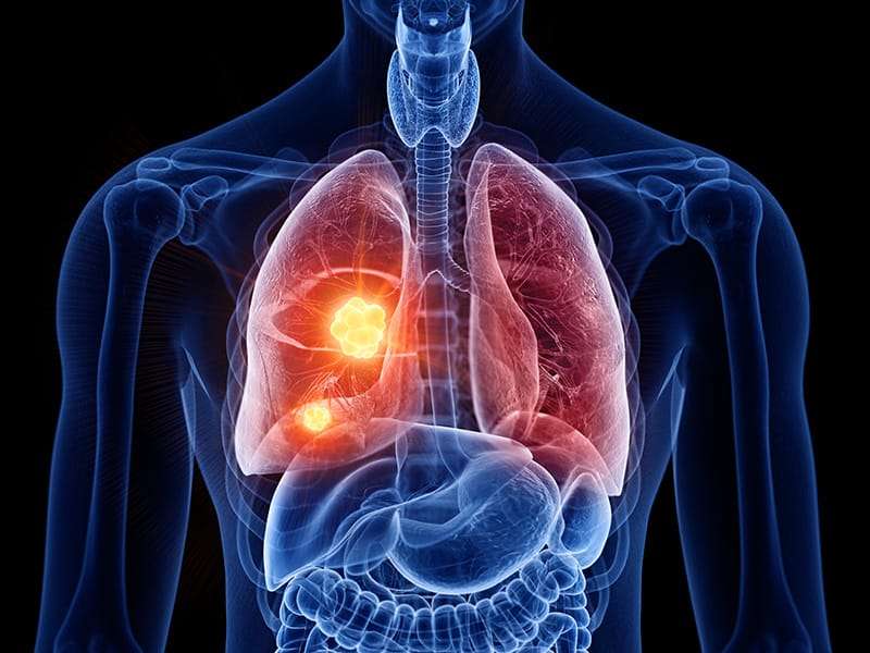
Lung surgery, also known as thoracic surgery, refers to surgical procedures performed on the lungs or other structures within the thoracic cavity. Lung surgery is often used to diagnose and treat various lung conditions, including lung cancer, lung infections, lung nodules, and certain lung diseases.
Lung surgery may be recommended for several reasons, including the removal of lung tumors (benign or malignant), treatment of lung infections (such as abscesses or empyema), management of lung conditions like bullae or lung cysts, and treatment of certain lung diseases like lung volume reduction surgery for advanced emphysema.
Prior to lung surgery, a thorough evaluation is conducted to assess the patient's overall health, lung function, and the extent of the lung condition. This evaluation may involve imaging studies (such as CT scans or PET scans), pulmonary function tests, blood work, and biopsies to determine the precise nature of any lung masses or nodules.
The surgical approach and incision depend on the specific procedure being performed. Traditional open surgery involves making an incision in the chest wall, often in the area between the ribs. Minimally invasive techniques, such as video-assisted thoracoscopic surgery (VATS), may be used, which involve smaller incisions and the use of specialized instruments and a video camera to access and visualize the lungs.
The specific surgical procedure depends on the underlying lung condition. Common lung surgeries include:
- Lobectomy: Removal of an entire lobe of the lung. This is often performed for lung cancer or certain benign lung conditions.
- Wedge Resection: Removal of a small, wedge-shaped portion of lung tissue. This may be performed for localized lung tumors or to remove lung nodules.
- Pneumonectomy: Removal of an entire lung. This is typically reserved for extensive lung tumors or in cases where one lung is severely damaged or infected.
- Segmentectomy: Removal of a specific segment of the lung. This procedure is performed when removing an entire lobe is not necessary, typically for smaller lung tumors or localized lung disease.
- Pleurodesis: A procedure to treat recurrent pleural effusion (fluid accumulation between the lungs and chest wall). It involves creating adhesions between the lung and chest wall to prevent fluid buildup.
Following lung surgery, patients typically require regular follow-up visits with their healthcare team. Rehabilitation and respiratory therapy may be recommended to aid in recovery and improve lung function. This may involve breathing exercises, physical activity, and guidance on lifestyle modifications to promote lung health.
It is important to note that lung surgery is a complex procedure and carries potential risks and complications, including bleeding, infection, blood clots, respiratory issues, and reactions to anesthesia. The specific approach and procedure chosen depend on the patient's condition, overall health, and the expertise of the surgical team. The decision to undergo lung surgery is made on an individual basis, weighing the potential benefits against the risks, and considering other
HEART VALVE REPAIR OR REPLACEMENT
Heart valve repair or replacement is a surgical procedure performed to treat heart valve diseases that involve damage or dysfunction of one or more of the heart's valves. The procedure aims to restore proper valve function and improve overall heart function.
Here's an overview of heart valve repair or replacement:
Heart valve repair or replacement may be recommended for various conditions, including valvular stenosis (narrowing of the valve) or regurgitation (leakage of blood backward through the valve). Common valve conditions that may require intervention include aortic valve stenosis or regurgitation, mitral valve regurgitation or stenosis, tricuspid valve regurgitation, and pulmonic valve stenosis or regurgitation.
Before the procedure, a comprehensive evaluation is conducted, which may include echocardiography, cardiac catheterization, stress tests, and other imaging studies to assess the valve's structure and function, determine the severity of the disease, and evaluate overall heart function.
The surgeon evaluates the valve and determines whether repair or replacement is necessary. Valve repair involves techniques to preserve and restore the natural valve structure, such as removing excess tissue, repairing the valve leaflets, or reinforcing the valve with a prosthetic ring. Valve replacement involves removing the damaged valve and replacing it with a mechanical valve (made of synthetic materials) or a biological valve (made from human or animal tissue).
After repairing or replacing the valve, the surgeon tests the valve's function and closes the incisions. Temporary pacing wires may be left in place to monitor the heart's electrical activity during the recovery period.
Following the procedure, the patient is transferred to the intensive care unit (ICU) for close monitoring. Over the next few days, the patient gradually transitions to a regular hospital room. Physical therapy, breathing exercises, and medications are provided to promote healing and recovery.
The patient may need to take blood-thinning medications (in the case of mechanical valve replacement) or other medications to prevent infection or manage symptoms. Heart valve repair or replacement is a major surgical procedure, and like any surgery, it carries risks such as bleeding, infection, blood clots, or complications associated with anesthesia.
CORONARY ARTERY BYPASS GRAFTING (CABG)
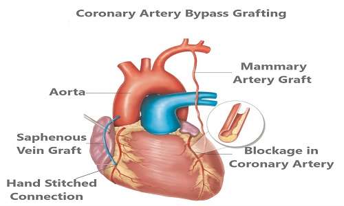
Coronary Artery Bypass Grafting (CABG) is a surgical procedure used to treat coronary artery disease (CAD), a condition characterized by the narrowing or blockage of the coronary arteries that supply blood to the heart muscle. CABG aims to restore normal blood flow to the heart by bypassing the blocked or narrowed arteries using grafts from other blood vessels.
Here's an overview of the CABG procedure:
1. Indications: CABG is typically recommended for individuals with severe coronary artery disease who experience symptoms such as chest pain (angina) or those who are at high risk of a heart attack. It is also considered for individuals with complex coronary artery blockages or when other treatments, such as medications or angioplasty, are not sufficient.
2. Preoperative Evaluation: Before undergoing CABG, a thorough evaluation is performed to assess the extent of coronary artery disease, heart function, and overall health. This evaluation may include tests such as angiography, stress tests, echocardiography, and blood work.
3. Anesthesia: CABG is performed under general anesthesia, meaning the patient is unconscious during the procedure. The anesthesiologist closely monitors vital signs and administers medications to ensure comfort and safety.
4. Incision and Exposure: The surgeon makes an incision in the middle of the chest (median sternotomy) to access the heart. Alternatively, minimally invasive techniques may be used, such as small incisions between the ribs (minithoracotomy) or robotic-assisted surgery.
5. Harvesting Grafts: Blood vessels are harvested from other parts of the body, typically the internal mammary artery from the chest wall and/or veins (saphenous veins) from the leg. These grafts will be used to bypass the blocked or narrowed coronary arteries.
6. Cardiopulmonary Bypass: To allow the surgeon to work on the heart, a heart-lung bypass machine is connected to the patient. The machine takes over the function of the heart and lungs, circulating and oxygenating blood throughout the body while the surgeon performs the bypass procedure.
7. Grafting: The surgeon carefully attaches the grafts to the coronary arteries, bypassing the blockages. The grafts serve as new pathways for blood to flow to the heart muscle, bypassing the diseased segments of the coronary arteries.
8. Conclusion: After completing the grafting, the surgeon verifies the grafts' function and closes the incisions. Temporary pacing wires may be left in place to monitor the heart's electrical activity during the recovery period.
9. Recovery: Following the procedure, the patient is transferred to the intensive care unit (ICU) for close monitoring. Over the next few days, the patient gradually transitions to a regular hospital room. Physical therapy, breathing exercises, and medications are provided to promote healing and recovery.
CABG is a major surgical procedure and, like any surgery, carries risks such as bleeding, infection, blood clots, and complications associated with anesthesia. However, it is a well-established procedure with a high success rate in relieving angina symptoms, improving quality of life, and prolonging survival in appropriate patients with severe coronary artery disease.
SKIN DISEASES
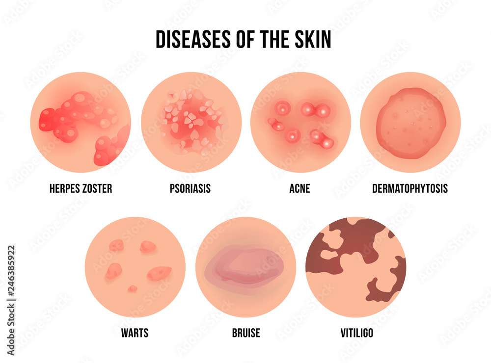
Skin diseases encompass a wide range of conditions that affect the skin, resulting in various symptoms and appearances. Some common skin diseases include:
- Acne: A common skin condition characterized by the formation of pimples, blackheads, and whiteheads on the face, chest, and back.
- Eczema: A chronic inflammatory condition that causes dry, itchy, and red patches on the skin.
- Psoriasis: A chronic autoimmune disease that causes red, scaly patches on the skin, often accompanied by itching or pain.
- Dermatitis: Inflammation of the skin that can be caused by various factors such as irritants, allergens, or genetic predisposition.
- Rosacea: A chronic skin condition characterized by facial redness, flushing, and sometimes pimples or visible blood vessels.
- Hives (Urticaria): Raised, itchy bumps on the skin that are often caused by an allergic reaction or exposure to certain triggers.
- Athlete's Foot: A fungal infection that affects the feet, causing itching, redness, and peeling of the skin.
- Ringworm: A fungal infection that forms a ring-shaped rash on the skin, often itchy and scaly.
- Impetigo: A highly contagious bacterial infection characterized by red sores that break open, ooze, and form a yellowish crust.
- Shingles: A viral infection caused by the reactivation of the varicella-zoster virus, resulting in a painful rash with fluid-filled blisters.
It's important to consult a dermatologist or healthcare professional for an accurate diagnosis and appropriate treatment for any skin condition.
CHILDHOOD DISEASES
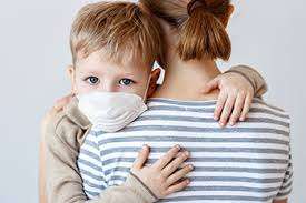
Childhood diseases refer to a wide range of illnesses that primarily affect children. Some common childhood diseases include:
- Chickenpox (Varicella): A highly contagious viral infection causing an itchy rash and fever.
- Measles: A viral infection characterized by fever, cough, runny nose, and a rash.
- Mumps: A viral infection causing swelling and pain in the salivary glands, leading to swollen cheeks and jaw.
- Rubella (German Measles): A viral infection causing a rash and fever, particularly dangerous for pregnant women.
- Whooping cough (Pertussis): A bacterial infection causing severe coughing spells, especially dangerous for infants.
- Hand, Foot, and Mouth Disease: A viral infection characterized by sores in the mouth, on the hands, and on the feet.
- Rotavirus: A highly contagious virus causing severe diarrhea, vomiting, and dehydration in infants and young children.
- Fifth Disease (Erythema infectiosum): A viral infection causing a distinctive rash on the face, cheeks, and body.
- Croup: A viral infection causing a barking cough and difficulty breathing.
- Roseola: A viral infection characterized by high fever followed by a rash.
It's important to note that diagnosing and treating specific diseases should be done by medical professionals. If you have concerns about a child's health, it's best to consult a healthcare provider.
WOMAN DISEASES
"Woman Disease" is a broad term that does not refer to a specific medical condition. Women can be affected by a variety of diseases and health conditions, some of which are unique to or more common in females. It's essential to address specific diseases or conditions to provide accurate information.
Here are some common health conditions that can affect women:
- Breast Cancer: A malignant tumor that develops in breast tissue, predominantly affecting women.
- Ovarian Cancer: Cancer that begins in the ovaries, the female reproductive organs responsible for producing eggs.
- Endometriosis: A condition where tissue similar to the lining of the uterus grows outside the uterus, causing pain and fertility problems.
- Polycystic Ovary Syndrome (PCOS): A hormonal disorder characterized by enlarged ovaries with cysts and hormonal imbalances, leading to irregular periods and other symptoms.
- Cervical Cancer: Cancer that develops in the cervix, the lower part of the uterus.
- Premenstrual Syndrome (PMS): A combination of physical and emotional symptoms that occur in the days leading up to menstruation.
- Urinary Tract Infections (UTIs): Infections in the urinary system, including the bladder and urethra, which can be more common in women.
- Osteoporosis: A condition characterized by a decrease in bone density, making bones more fragile and prone to fractures, which is more common in women, especially after menopause.
- Menopause: The natural process that occurs in women's lives, typically in their 40s or 50s when menstrual periods cease and hormone levels change.
- Pelvic Inflammatory Disease (PID): An infection of the female reproductive organs, usually caused by sexually transmitted bacteria.
It's important to note that these are just a few examples, and there are many more health conditions that can affect women. If you have concerns about your health, it's recommended to consult a healthcare professional for a proper diagnosis and treatment.
VACCINATION AND CHECK-UP
INFECTIOUS DISEASES

GASTRIC DISORDERS
PAIN MANAGEMENT

MIGRAINE

RESPIRATORY PROBLEMS
HEART PROBLEMS
HEART PROBLEMS

THYROID DISEASE
DIABETES MANAGEMENT

SORE THROAT