
Dr Griz Gupta
About Me
Dr Griz Gupta
MS
Orthopaedic Surgeons
10 Years Experience
Biography
My Clinic
OPD Hours
| Monday | Morning | 10:00 AM | - | 02:00 PM |
| Evening | 06:00 PM | - | 09:00 PM | |
| Tuesday | Morning | 10:00 AM | - | 02:00 PM |
| Evening | 06:00 PM | - | 09:00 PM | |
| Wednesday | Morning | 10:00 AM | - | 02:00 PM |
| Evening | 06:00 PM | - | 09:00 PM | |
| Thursday | Morning | 10:00 AM | - | 02:00 PM |
| Evening | 06:00 PM | - | 09:00 PM | |
| Friday | Morning | 10:00 AM | - | 02:00 PM |
| Evening | 06:00 PM | - | 09:00 PM | |
| Saturday | Morning | 10:00 AM | - | 02:00 PM |
| Evening | 06:00 PM | - | 09:00 PM | |
| Sunday | Morning | 10:00 AM | - | 02:00 PM |
| Evening | 06:00 PM | - | 09:00 PM |
Treatments
Specialised care and treatments offered
ELECTROMYOGRAPHY (EMG) FOR NERVE ASSESSMENT
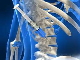
Electromyography (EMG) assesses nerve function by measuring electrical activity generated during muscle contractions. This diagnostic tool is valuable in identifying neuromuscular disorders affecting muscle control due to nerve damage or dysfunctions.
. < !DOCTYPE HTML PUBLIC " -//W3C//DTD HTML 4.01 Transitional//EN" "http://www.w3.org/TR/html4/loose.dtd">{ . -{/>}& archive .= URL's + & &.[ - +P(d T )]}
- Symptoms : Patients might report tingling sensations, weakness in limbs ,and difficulty performing everyday tasks indicating potential nerve involvement .
- Diagnostic Procedure : During EMG testing electrodes placed on skin record electrical signals elicited during voluntary muscle movements , providing critical data regarding muscular health status .
- Treatment Guidance : Understanding test results supports clinicians deciding whether rehabilitative therapies ,medicinal approaches ,or surgical options would benefit patient outcomes after defining underlying issues clearly .
- < span style="font-weight:bold;">•< span style="font-weight:bold;">…< b />< ul />~< p /> Nerve assessments through EMG serve as an integral part aiding healthcare providers navigate complex cases involving debilitating conditions impacting functional independence swiftly!.< p/>
BONE DENSITY TESTING (DEXA SCAN)

A Dual-Energy X-ray Absorptiometry (DEXA) scan measures bone mineral density (BMD) and evaluates fracture risk due to conditions like osteoporosis. This vital test helps identify weakened bones before they break or fracture under pressure applied during everyday activities.
- Risk Factors : People experiencing chronic steroid use or loss of height may require further investigation by DEXA scanning.
- Diagnosis Process : Technicians position patients comfortably while performing scans lasting about 10–30 minutes depending upon coverage areas requested.
- Treatment Implications : If low BMD values emerge from tests signifying osteoporosis presence additional lifestyle modifications alongside medications could be required. < ~ * & ~*.*~ # ~ * . “I‘ ”{ –{ ;``--’-’ ;}
Bone density testing serves as an essential preventive measure helping individuals comprehend their skeletal health status while aiding practitioners develop proactive approaches toward maintaining optimal bone strength throughout one's lifetime!.
ULTRASOUND FOR SOFT TISSUE EVALUATION

Ultrasound imaging employs sound waves to produce live images of soft tissues within the body. It is especially effective in evaluating conditions affecting muscles, tendons, and ligaments around joints. This technique is safe and does not involve radiation exposure.
- Candidates for Ultrasound: Patients with unexplained pain or stiffness often benefit from ultrasound assessments that visualize soft tissue structures comprehensively.
- How It Works: A specially trained sonographer applies a gel on the skin over the area of interest followed by using a transducer that emits sound waves to create real-time images on a monitor.
- Treatment Insights: The information gathered can inform management strategies; whether it involves rest and rehabilitation or potential minimally invasive surgeries if tears are identified within tendons or ligaments.
The immediacy of ultrasound results allows healthcare professionals to make prompt decisions regarding treatment plans while reassuring patients through its non-invasive nature!
CT SCANS FOR COMPLEX INJURIES

Computed Tomography (CT) scans offer detailed cross-sectional images of bones and soft tissues, which makes them particularly useful in diagnosing complex orthopedic injuries. This advanced imaging technique can uncover subtler details that traditional X-rays might miss.
- Symptoms Indicating Need: Intense pain following trauma or chronic joint issues may lead physicians to recommend a CT scan for deeper insights.
- Diagnostic Approach: During this test, multiple X-ray images are taken from different angles, which are then compiled into comprehensive 3D models by special software.
- Treatment Outcomes: Results guide doctors in recommending specific interventions—non-surgical methods like physiotherapy or necessary surgical procedures when significant damage is present.
- Results Overview: Most people feel comfortable during CT scans as they require minimal movement, resulting in high-quality images essential for accurate assessment.
This state-of-the-art diagnostic tool significantly enhances decision-making processes within orthopedic practice when managing both acute injuries and chronic conditions effectively.
X-RAYS FOR BONE FRACTURES

X-rays are among the most common diagnostic tools used to detect bone fractures. This imaging method uses radiation to create images that reveal broken bones or other abnormalities. It's a fast and efficient way to assess injuries sustained from accidents or falls.
- Symptoms: Common signs indicating a fracture can include intense pain, swelling, and deformity in the affected area.
- How It’s Diagnosed: A doctor will examine the injury before taking X-rays to confirm the fracture type and its location.
- Treatment Options: Treatment might range from casting and bracing minor fractures to surgical intervention for more severe breaks requiring fixation.
- Aftercare: Post-diagnosis care is essential; follow-up visits ensure proper healing and realignment if needed.
X-ray imaging remains a fundamental aspect of orthopedic practices, allowing for quick diagnosis and timely intervention in cases of suspected fractures.
MRI SCANS FOR JOINT ISSUES

Magnetic Resonance Imaging (MRI) scans are a non-invasive imaging technique used to visualize internal structures of the body, particularly useful in assessing joint problems. They provide detailed images of soft tissues, including cartilage and ligaments, making them invaluable in the diagnosis of various orthopedic conditions.
- Indications: Patients may experience persistent joint pain, swelling, or limited mobility, prompting an MRI.
- Diagnostic Process: An MRI technician positions the patient inside the scanner, where they must remain still for accurate imaging.
- Treatment Options: Depending on findings, treatments may include physical therapy or surgical interventions if severe damage is detected.
- What to Expect: The procedure is painless and typically lasts 30-60 minutes; afterwards, patients can resume normal activities.
MRI scans play a crucial role in informing treatment decisions for joint-related issues, ensuring patients receive tailored care plans based on precise diagnoses.