
Dr Gauri Shankar
About Me
Dr Gauri Shankar
MS
Orthopaedic Surgeons
10 Years Experience
Biography
My Clinic
OPD Hours
| Monday | Morning | 10:00 AM | - | 02:00 PM |
| Evening | 06:00 PM | - | 09:00 PM | |
| Tuesday | Morning | 10:00 AM | - | 02:00 PM |
| Evening | 06:00 PM | - | 09:00 PM | |
| Wednesday | Morning | 10:00 AM | - | 02:00 PM |
| Evening | 06:00 PM | - | 09:00 PM | |
| Thursday | Morning | 10:00 AM | - | 02:00 PM |
| Evening | 06:00 PM | - | 09:00 PM | |
| Friday | Morning | 10:00 AM | - | 02:00 PM |
| Evening | 06:00 PM | - | 09:00 PM | |
| Saturday | Morning | 10:00 AM | - | 02:00 PM |
| Evening | 06:00 PM | - | 09:00 PM | |
| Sunday | Morning | 10:00 AM | - | 02:00 PM |
| Evening | 06:00 PM | - | 09:00 PM |
Treatments
Specialised care and treatments offered
DISPLACED DISTAL RADIUS FRACTURE
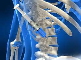
This type of wrist fracture occurs near the end of the radius bone when it breaks with fragments shifting out of normal alignment. Commonly seen in falls onto an outstretched hand especially among active individuals or elderly falling accidents . Proper alignment is critical in recovering wrist function fully .
- Patients experience wrist pain , swelling , deformity resembling a 'dinner fork' contour , limited wrist movement immediately post-injury .
- Diagnosis involves detailed physical exam supported primarily by X-rays confirming displacement degree & comminution pattern helping guide treatment planning .
- Management ranges from closed reduction casting in stable cases through ORIF (open reduction internal fixation) surgery using plates & screws for unstable displaced fragments restoring proper anatomical position , maintaining joint congruence & enabling early rehabilitation sessions improving grip strength & dexterity recovery .
- Post-treatment expectations include gradual symptom relief over weeks-months coupled with persistent physiotherapy maximizing range-of-motion , preventing stiffness .
Careful treatment decisions ensure satisfactory functional outcomes minimizing chronic pain risk associated complications involving distal radius fractures .
FEMORAL SHAFT FRACTURE
A femoral shaft fracture involves a break along the long straight part of the thighbone. Typically resulting from high-energy trauma such as vehicle accidents or falls from height, this injury can cause significant disability without prompt care.
- This fracture presents with intense thigh pain deformity, inability to bear weight on the leg, swelling, bruising, and sometimes visible shortening of limb length compared to the other side.
- X-rays confirm fracture location/type; occasionally CT scans provide additional detail if complex anatomy is involved.
- Surgical fixation using intramedullary nails or plates usually provides stable internal support allowing early mobilization while promoting bone union effectively over weeks/months post-op period involving physiotherapy aim at muscle strengthening & joint flexibility restoration once stable healing achieved .
Timely surgery combined with rehabilitation optimizes return to normal function following femoral shaft fractures especially after major trauma scenarios .
ACUTE ACHILLES TENDON RUPTURE

The Achilles tendon connects calf muscles to the heel bone; a sudden rupture during sports or strenuous activity causes marked impairment in walking or standing on tiptoes. Immediate care is vital for recovery.
- Patients often hear a popping sound followed by sharp pain near the heel alongside swelling or bruising behind the ankle.
- The diagnosis relies on clinical tests like Thompson’s test supported by ultrasound or MRI imaging if needed.
- Treatment options vary between conservative methods using casting or braces versus surgical repair depending on patient age, activity level, and rupture severity.
- Rehabilitation programs focus on gradual weight-bearing restoration along with stretching & strengthening exercises tailored individually for optimal healing outcomes.
An informed approach improves functional results by balancing healing time with early mobilization benefits after an Achilles tendon rupture event.
TRAUMATIC SHOULDER DISLOCATION

A traumatic shoulder dislocation occurs when the upper arm bone pops out of its socket due to sudden impact or forceful movement. It can significantly limit arm mobility and cause acute pain.
- The most obvious symptom is a visibly deformed shoulder accompanied by intense discomfort and swelling immediately after injury.
- A physical examination combined with imaging such as X-rays confirms the diagnosis and rules out associated fractures.
- Treatment typically begins with closed reduction — manually repositioning the humerus into its proper place under guided care.
- This is followed by immobilization in a sling for several weeks alongside pain management strategies and rehabilitation exercises focusing on strengthening shoulder muscles.
Early treatment promotes better joint stability while reducing chances of recurrent dislocations or chronic instability issues later on.
COMPARTMENT SYNDROME

Compartment syndrome arises when excessive pressure builds within muscle compartments, compromising blood flow and tissue viability. This condition often follows trauma or fractures in limbs and demands urgent attention.
- Signs include intense pain disproportionate to injury, numbness or tingling, swelling, and decreased limb function.
- The diagnosis is primarily clinical but can be aided by measuring intracompartmental pressures.
- Treatment is emergent, commonly requiring a surgical procedure called fasciotomy to relieve pressure within affected compartments.
- If untreated, it may lead to permanent muscle and nerve damage; thus rapid recognition is essential.
A swift response with appropriate intervention can prevent lasting disability from compartment syndrome by restoring circulation promptly.
OPEN FRACTURE MANAGEMENT

Open fractures are serious injuries where the broken bone pierces the skin, exposing the fracture to the external environment. Prompt and effective treatment is crucial to prevent complications such as infections and improper healing.
- Symptoms include visible bone protrusion, bleeding, severe pain, and swelling around the injury site.
- Diagnosis typically involves a physical exam and imaging tests like X-rays to evaluate the extent of bone damage and soft tissue involvement.
- Treatment requires immediate wound cleaning, stabilization with splints or surgical fixation, and administration of antibiotics to reduce infection risk.
- Surgical intervention might be necessary to align bones, remove dead tissue, and close wounds appropriately while preserving blood supply.
- Recovery focuses on careful monitoring for infection signs, physical therapy for restoring function, and regular follow-up imaging.
Timely management of open fractures substantially improves outcomes by minimizing infection risks and promoting optimal healing of both bone and soft tissues.