
Dr Abhishek Agar
About Me
Dr Abhishek Agar
MBBS
Ophthalmologist
10 Years Experience
Biography
My Clinic
OPD Hours
| Monday | Morning | 10:00 AM | - | 02:00 PM |
| Evening | 06:00 PM | - | 09:00 PM | |
| Tuesday | Morning | 10:00 AM | - | 02:00 PM |
| Evening | 06:00 PM | - | 09:00 PM | |
| Wednesday | Morning | 10:00 AM | - | 02:00 PM |
| Evening | 06:00 PM | - | 09:00 PM | |
| Thursday | Morning | 10:00 AM | - | 02:00 PM |
| Evening | 06:00 PM | - | 09:00 PM | |
| Friday | Morning | 10:00 AM | - | 02:00 PM |
| Evening | 06:00 PM | - | 09:00 PM | |
| Saturday | Morning | 10:00 AM | - | 02:00 PM |
| Evening | 06:00 PM | - | 09:00 PM | |
| Sunday | Morning | 10:00 AM | - | 02:00 PM |
| Evening | 06:00 PM | - | 09:00 PM |
Treatments
Specialised care and treatments offered
YAG LASER CAPSULOTOMY
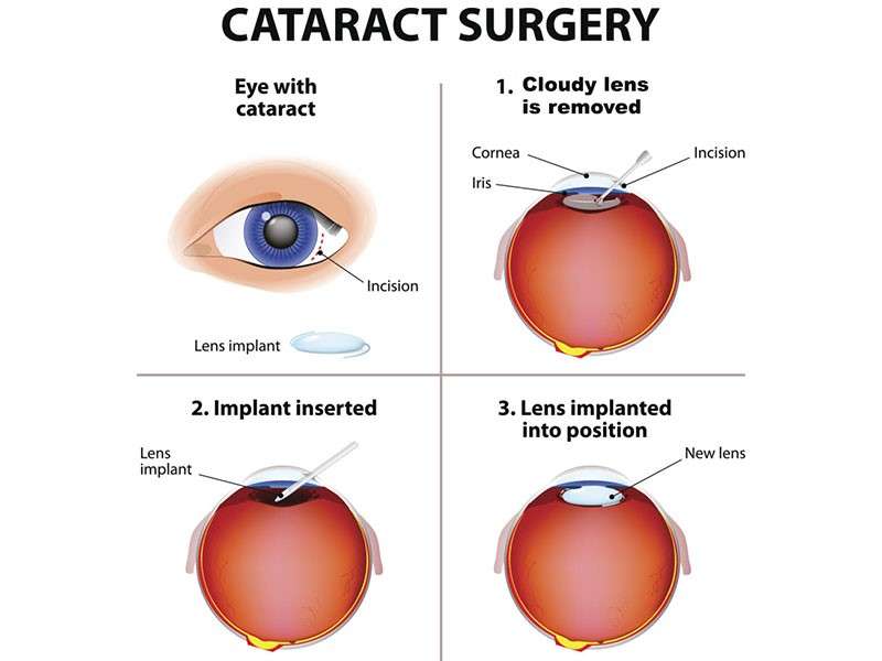
The YAG laser capsulotomy treats posterior capsule opacification (PCO), a frequent delayed complication after cataract surgery causing cloudy vision akin to original cataract symptoms. This quick procedure restores clarity efficiently without incisions.
- Nature of PCO: Opacification develops behind implanted lens months or even years post-cataract operation affecting visual acuity.
- Identification: Patients report blurred images or glare worsening despite prior successful surgery; slit lamp confirms diagnosis.
- Treatment Method: A focused YAG laser creates an opening in the opaque capsule allowing unobstructed light passage through pupil.
- Procedure Benefits: Performed outpatient without anesthesia painlessly; immediate improvement typically observed.
This minimally invasive intervention revitalizes quality of sight helping individuals regain comfortable daily functioning quickly after cataract care completion.
PTERYGIUM REMOVAL

A pterygium is a benign growth of conjunctival tissue over the cornea often related to chronic sun exposure. Surgical removal can relieve irritation and improve appearance when it causes symptoms or threatens vision clarity.
- Recognition: Symptoms include redness, dryness, foreign body sensation, and visual distortion if growth encroaches on central cornea.
- Diagnosis: Clinical examination by slit lamp confirms extent and vascularity of lesion.
- Surgical Intervention: Excision usually followed by conjunctival autograft placement reduces recurrence rates significantly.
- Postoperative Expectations: Mild discomfort controlled by medications; visual recovery occurs over weeks.
Proper preventive measures like UV protection alongside timely treatment ensure long-term ocular surface health against this common outdoor-related condition.
LASER TREATMENT FOR DIABETIC RETINOPATHY

This advanced laser treatment addresses damage caused by uncontrolled diabetes affecting retinal blood vessels. It aims to reduce risk of severe vision loss from diabetic retinopathy complications.
- Condition Details: Diabetic retinopathy involves leakage or abnormal growth of blood vessels within the retina due to prolonged high blood sugar levels.
- Treatment Approach: Laser photocoagulation seals leaking vessels and reduces abnormal new vessel formation thereby stabilizing retina health.
- Procedure Format: Performed on an outpatient basis under local anesthesia with precise targeting using specialized lasers conserving surrounding tissue.
- Follow-up Care: Multiple sessions may be required; regular retinal exams monitor therapy response and detect recurrence early on.
This technique plays a vital role in preserving sight among diabetic patients when combined with systemic glucose control strategies.
GLAUCOMA EVALUATION & MANAGEMENT

Glaucoma refers to a group of conditions causing optic nerve damage typically due to increased intraocular pressure. Early detection is crucial as it prevents irreversible vision loss.
- Symptoms: Often asymptomatic in early stages; advanced disease may cause peripheral vision loss or tunnel vision.
- Diagnostic Tools: Tonometry to measure eye pressure, optic nerve imaging, visual field testing are key components of evaluation.
- Treatment Modalities: Use of medicated eye drops to lower eye pressure; laser therapy or minimally invasive surgeries are options when medications are insufficient.
- Lifelong Monitoring: Regular check-ups are essential since glaucoma requires ongoing management to preserve vision.
Timely intervention can control progression effectively and keep daily activities safe for patients living with glaucoma.
CATARACT SURGERY
Cataracts cause clouding of the natural lens inside the eye, leading to blurry vision that progressively worsens. While surgery is often required to restore sight fully, advances have made it safer and quicker than ever.
- Symptoms: Blurred or dim vision, difficulty with night vision, sensitivity to glare, and seeing halos around lights.
- Diagnosis: Detailed eye exam including slit-lamp microscopy to assess lens opacity.
- Surgical Procedure: Minimally invasive phacoemulsification involves breaking up the cloudy lens with ultrasound and implanting a clear intraocular lens (IOL).
- Recovery & Outcomes: Most patients experience rapid visual improvement with minimal discomfort within days after surgery.
This procedure is one of the most commonly performed surgeries worldwide due to its effectiveness in restoring clear vision safely.
DRY EYE SYNDROME
Dry Eye Syndrome is a common condition where the eyes do not produce enough tears or the tears evaporate too quickly, leading to discomfort and vision issues. It can affect people of all ages and may worsen over time if left untreated.
- Symptoms: Irritation, burning sensation, redness, and a feeling of grittiness or foreign body sensation in the eyes.
- Causes: Aging, environmental factors such as wind or dry climates, prolonged screen time, and certain medications.
- Diagnosis: Comprehensive eye examination including tear film tests like Schirmer's test or tear breakup time assessment.
- Treatment Options: Artificial tears, lifestyle changes such as reducing screen exposure, warm compresses, and sometimes prescribing anti-inflammatory eye drops.
- Expectations: Proper management can significantly improve comfort and prevent further damage to the ocular surface.
Early identification and consistent treatment help maintain optimal eye health and improve quality of life for those affected by this prevalent condition.