
Dr A K Aggarwal H
About Me
Dr A K Aggarwal H
MBBS
Cardiologist
10 Years Experience
Biography
My Clinic
OPD Hours
| Monday | Morning | 10:00 AM | - | 02:00 PM |
| Evening | 06:00 PM | - | 09:00 PM | |
| Tuesday | Morning | 10:00 AM | - | 02:00 PM |
| Evening | 06:00 PM | - | 09:00 PM | |
| Wednesday | Morning | 10:00 AM | - | 02:00 PM |
| Evening | 06:00 PM | - | 09:00 PM | |
| Thursday | Morning | 10:00 AM | - | 02:00 PM |
| Evening | 06:00 PM | - | 09:00 PM | |
| Friday | Morning | 10:00 AM | - | 02:00 PM |
| Evening | 06:00 PM | - | 09:00 PM | |
| Saturday | Morning | 10:00 AM | - | 02:00 PM |
| Evening | 06:00 PM | - | 09:00 PM | |
| Sunday | Morning | 10:00 AM | - | 02:00 PM |
| Evening | 06:00 PM | - | 09:00 PM |
Treatments
Specialised care and treatments offered
BLOOD PRESSURE MONITORING

The assessment of blood pressure regularly allows early identification and management of hypertension which remains a leading cause of cardiovascular morbidity worldwide. Proper monitoring helps reduce risks associated with untreated high blood pressure such as stroke and heart failure.
- Purpose: Detect elevated blood pressure readings over repeated measurements necessitating lifestyle modifications or medications.
- Techniques: Available methods include office-based measurements using sphygmomanometers as well as ambulatory devices providing continuous data across day-night cycles.
- Clinical Impact: Consistent monitoring identifies uncontrolled hypertension despite therapy enabling timely treatment adjustments minimizing complications.
- Patient Role: Encourages active engagement via home blood pressure tracking improving compliance with prescribed interventions.
Sustained control through vigilant monitoring significantly decreases long-term adverse effects promoting healthier cardiovascular outcomes overall.
CARDIAC CATHETERIZATION
This invasive diagnostic procedure provides direct visualization and measurement of pressures inside coronary arteries and cardiac chambers. Cardiac catheterization is fundamental when non-invasive tests suggest significant disease needing confirmation or intervention.
- Pain Control & Preparation:The procedure involves numbness at insertion sites typically through wrist or groin arteries accompanied by sedation if necessary so patient comfort is ensured throughout test duration lasting roughly an hour or two.
- Diagnostic Goals: Pinpoints degree/location of arterial blockages causing angina or myocardial infarction risk while assessing ventricular performance precisely.
- Treatment Integration: Often combined with angioplasty/stenting immediately if critical stenosis detected avoiding need for separate surgery.
- Post-procedure Monitoring: Observation follows intervention period ensuring vascular access site healing plus complication prevention.
Considered gold-standard test when pinpoint diagnosis followed by immediate therapy may improve survival rate considerably among advanced coronary artery disease cases.
HOLTER MONITORING
A Holter monitor is a portable device worn by patients temporarily to track continuous ECG recordings over 24 to 48 hours. It captures intermittent arrhythmias that might be missed during short clinic visits.
- Sensitivity Advantage: Ideal for detecting occasional palpitations or unexplained fainting spells related to abnormal rhythms.
- User Experience: Patients carry on regular activities while wearing small electrodes connected to the lightweight monitor attached around the waist or shoulder strap area.
- Description of Data Collection:Create detailed logs correlating symptoms with recorded electrical events of the heart throughout daily life routines.
- Treatment Impact:The collected data assists doctors in determining specific arrhythmia types needing medication adjustment or intervention procedures like pacemaker implantation.
This ambulatory monitoring significantly improves diagnostic accuracy beyond standard ECG snapshots taken at rest in clinical environments.
STRESS TESTING (EXERCISE TOLERANCE TEST)
The Stress Test evaluates how well your heart functions during physical exertion. This test helps uncover coronary artery blockages not seen while at rest by monitoring cardiac performance under stress.
- Main Use: Identifies symptoms triggered by exercise such as chest discomfort or shortness of breath linked to reduced blood supply.
- Test Process: Patients walk on a treadmill or pedal a stationary bike while their ECG, blood pressure, and symptoms are monitored continuously.
- Result Insights: Abnormal changes indicate potential coronary artery disease requiring further treatment plans.
- Safety Measures: Conducted under medical supervision with readiness for emergency intervention if needed.
This procedure provides critical information about cardiovascular fitness levels and guides risk stratification for patients suspected of having ischemic heart disease.
ELECTROCARDIOGRAM (ECG)
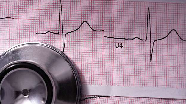
An Electrocardiogram (ECG) records the electrical activity of the heart over time. It is one of the most common tests used to detect irregularities in heartbeat patterns or rhythm disturbances.
- Symptoms Indicated: Useful for patients experiencing chest pain, palpitations, dizziness, or fainting episodes.
- How It's Performed: Small electrodes are placed on the skin of the chest, arms, and legs to measure electrical signals from the heart.
- Diagnoses Possible: Detects arrhythmias, myocardial infarction (heart attack), ischemia, and electrolyte imbalances.
- Interpretation: The test results guide further investigations or therapeutic interventions based on abnormalities detected.
The ECG test is quick, painless, and critical for evaluating cardiac electrical function in both emergency and routine settings.
ECHOCARDIOGRAPHY
Echocardiography is a vital imaging technique that uses sound waves to produce detailed images of the heart's structure and function. It helps physicians assess heart health, detect abnormalities, and plan appropriate treatments.
- Purpose: Identifies issues like valve problems, chamber size changes, and heart muscle function.
- Procedure: A non-invasive test where a transducer emits ultrasound waves onto the chest wall to capture moving images of the heart.
- Diagnostic Benefits: Assists in diagnosing cardiomyopathies, congenital heart diseases, and pericardial effusions.
- Patient Prep & Experience: Usually requires no special preparation; takes about 30-60 minutes and is painless.
- Outcome: Provides comprehensive insight into cardiac health for effective management decisions.
This method offers a safe and informative way to evaluate heart conditions without exposure to radiation, making it an essential tool in cardiology diagnostics.